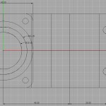
Update: here is the plate. Let's hope it is accurate enough (it is not face-milled on the underside...)
A plate for holding 76 mm x 26 mm glass slides in the microscope. My first ever 'real' drawing with LibreCAD (that website has been down for two days now, so try also librecad on sourceforge).
The fourth paper from my thesis, entitled "Dual-trap optical tweezers with real-time force clamp control", has just been published online by Review of Scientific Instruments: http://link.aip.org/link/doi/10.1063/1.3615309
Here's a video from the paper. We are holding on to two micron sized plastic spheres with laser-beams (shown in the video as green/cyan cross-hairs). The lower beam/trap is stationary while the upper one is steerable. A ca 16um long DNA-molecule (invisible) is tethered between the beads.The experiment is performed in the presence of lambda exonuclease, an enzyme that "eats up" one strand of the DNA leaving just a single-stranded DNA-tether between the beads.
In the first part of the video a force-extension curve (bottom panel) is obtained using manual control. We stretch out the molecule by moving the upper trap upwards and check that the force-signal looks like it should when we have a single DNA-molecule of the right length between the beads.
In the second part, after t = 20 s, the tether is held force clamped at 3.4 pN (force shown in top panel). We're keeping the force constant with a PI-controller implemented on an FPGA that reads the force-signal from the lower bead and updates the position of the upper trap at around 200 kHz. As the molecule shortens the controller needs to move the upper trap/bead lower in order to maintain a 3.4 pN tension in the molecule. The video is at normal speed (1X) while the force extension curve is measured. During 13 min of force-clamp control the video is sped up 25-fold. During this time the exonuclease digests one strand of the double-stranded DNA molecule. When held at 3.4 pN of tension, single-stranded DNA is significantly shorter than double-stranded DNA. So the gradual conversion from a double-stranded tether to a single-stranded tether is seen as a decrease in the extension, i.e. a shortening of the distance between the plastic beads (middle panel). The tether broke at t = 880 s. Scale-bar 5 ?m.
A ~16 um long DNA-molecule is tethered between optically trapped plastic beads. Beads are held by a stationary trap (lower blue cross-hairs) and a steerable trap (upper green cross-hairs). The graphs on the right show the measured force (red) and the force set-point (blue) (top), the distance between the traps (middle), and the force-extension curve with a green cross indicating the current value (bottom). A force-extension curve of the tether is first obtained manually, before force-clamp feedback is switched on at t=24 s. The force set-point is first at 5.5 pN, then increased to 11.4 pN at 30 s and finally increased to 17.4 pN at 35 s. Scale-bar 5 µm. Anders Wallin et al. University of Helsinki, Finland, 2011.
Some very early testing of fluorescence imaging in our optical tweezers instrument. A 10 kb long piece of DNA (ca 3 um long when stretched) is held between two optically trapped microspheres. The DNA is coated with a fluorescent dye (SYBR-gold) which is exited by a 488 nm blue laser and the fluorescence signal is collected with a CCD camera looking through a narrow-band filter centered on the emission spectrum of SYBR-gold.
At around 1:10 in the video there's a double-tether (two DNA-molecules between the beads). We don't want that but there is not much that we can do about it, except discard the data. At the very end there's an image of QDots on the coverglass surface.
Here a DNA-molecule is being stretched between two optically trapped polystyrene micron-sized beads. We're using an FPGA-based real-time controller for steering the upper trap. It's programmed with a PI-loop which aims to keep the force acting on the lower bead constant. Around 10s into the video we switch on the feedback-loop and we see the actual force on the bead rise to the set-point.
A ~48 000 base-pair long (ca 16 um) piece of DNA is stretched between two optically trapped ca 2 um diameter polystyrene beads. Bright-field real-time view through a 100x microscope. Scale-bar in microns on the right.
Were stretching a 13 kb DNA-construct between two optically trapped beads. This isn't a video of the experiment itself, but of some entangled beads we found in an old sample. The beads are 3 or 2 micron in diameter, and we hold on to the middle big bead with the optical trap while moving around the microscope stage. Hydrodynamic drag stretches out the chain of entangled beads. Not much, if any, scientific value, but a fun video nevertheless.
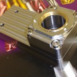
I'm fitting some more equipment around a Nikon TE-2000 microscope, and the stock objective turret is in the way. Machined this objective holder in steel yesterday.